Designing High-Quality Images for Research Papers and Theses: The Available Tools


Manuscripts express data collected from months or years of careful experimentation. However, raw data or narratives alone don’t make good journal articles. Data visualization tools and free drawing software enable scientists to explain their scientific story. By using tools to perfect scientific illustration, your manuscript can grab reviewers’ attention. More importantly, it will help your readers understand data quickly, increasing the likelihood of citing and sharing your research paper.
Why Image Quality Matters
Journals have strict guidelines regarding figure/image quality (e.g. “dots per inch”/DPI or number of pixels per image). Editors and their staff will turn down manuscripts prior to review if the images are of insufficient quality. Furthermore, poor figure quality can leave a bad impression on readers and reviewers. So, when editing and creating scientific images, be sure to use scientific illustration software or drawing tools to make your data clear and understandable!
Tables can help communicate data quickly to readers, who are often short on time. For this reason, when you have a well-designed table, your paper can have a far greater impact. For this reason, your tables should have clear and descriptive titles, well-defined headings, aligned data entries in each cell, and clearly defined units for all data entries. Meanwhile, when designing figures, there are many tools available to researchers to create publication-ready images.
Related: Creating images for your research paper? Check out these resources and avoid image manipulation now!
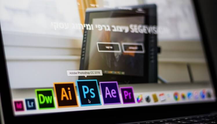
Uses and Limitations of Common Tools
There is a myriad of tools available for scientists. Picking which one to work with depends on your computer literacy, budget, and desired outcomes.
R is a free statistics computing program that also facilitates graphics development. It works on a variety of operating systems. Furthermore, the default design choices for image rendering were made to generate publication-quality plots with ease. While it is free, it is not as user-friendly as subscription services, such as Prism, which allows for both data analysis and figure development.
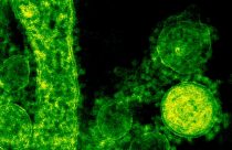
ImageJ is a freely available software developed by the National Institutes of Health. In short, it is an image-processing program that allows users to edit, analyze, process, save, print, modify colors, and quantitate images. One of the more exciting features is its ability to generate stacks (a series of images) from videos or convert photos into videos. This is helpful for live cell imaging.
Inkspace is a quality vector graphics editor that is open sourced and provides flexible drawing tools. It has broad file format compatibility and a powerful text tool.
GIMP is a free image manipulation program that can be combined with plugins to enhance features. It requires greater computer literacy than most other image formatting tools.
Cytospace is an open source network for complex network analysis that helps users integrate, analyze, and visualize data. While it is free to use, it is not as user-friendly as Ingenuity Pathway Analysis, which allows for pathways to be designed and rendered artistically with great ease (for a fee, of course).
ImageMagick is another tool that can be used to read and write images in many commonly used formats (e.g., PNG, JPEG, FIG, TIFF, PDF, etc). For this reason, it can modify images in nearly any manner. It allows users to composite images, animate, manage color, decorate, draw, and delineate image features (e.g., edges of colors). Furthermore, it is compatible with multiple coding languages.
Tips
- While creating figures can be a fun process, it’s important to always do it correctly. First, check the required format for images prior to submitting. If you have to convert the image file, check to ensure that your DPI is still at least 300.
- Once ready to submit, carefully review figures for errors prior to publishing. One method of doing this is to print your figures in color and review them manually. This will help you spot oddities that may have otherwise been missed by an electronic review.
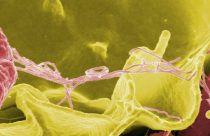
- When modifying your images for publication, never manipulate your images in a manner that is fraudulent. Western blots are often the most suspicious images available that will carefully be scrutinized by your reviewers.
- Finally, while a lot of data is helpful to have, be sure to reduce the presence of “chartjunk” – the unnecessary visual elements that distract the reader from what really matters…your data!